
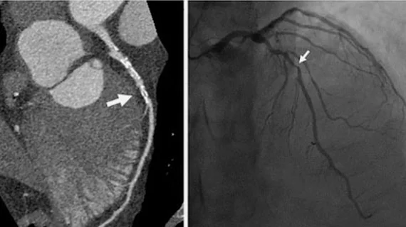
When comparing a cardiac CT scan vs angiogram, both diagnostic tests provide valuable insights into your heart’s health, but they work in different ways and are suited to different clinical situations. Understanding the differences between these two imaging techniques can help you feel more prepared for your consultation and treatment journey.
Dr Naveen Mudalagiri is an experienced award winning interventional cardiologist practising in Central London and Kent who specialises in advanced cardiac diagnostics and heart care. With expertise in both non-invasive and invasive cardiac procedures, Dr Mudalagiri helps patients understand which diagnostic approach may be most appropriate for their individual circumstances.
What Is a Cardiac CT Scan?
A cardiac CT scan, also known as a coronary CT angiogram or cardiac computed tomography, is a non-invasive imaging test that uses X-rays and computer technology to create detailed pictures of your heart and coronary arteries. This sophisticated diagnostic tool can reveal important information about your heart’s structure and blood vessels without requiring any invasive procedures.
During a cardiac CT scan, you lie on a table that slides into a doughnut-shaped scanner. The machine rotates around your body, taking multiple X-ray images from different angles. These images are then processed by a computer to create cross-sectional views of your heart and coronary arteries.
How Does Cardiac CT Scanning Work?
The cardiac CT scan process typically involves the administration of contrast dye through an intravenous line in your arm. This contrast material helps highlight your blood vessels and heart structures, making them more visible on the images. The entire scanning process usually takes between 10 to 30 minutes, though the actual image acquisition may only take a few seconds to a few minutes.
Modern CT scanners can capture images of your beating heart with remarkable clarity, allowing cardiologists to assess the coronary arteries for signs of narrowing, blockages, or other abnormalities that might affect blood flow to your heart muscle.
What Is an Angiogram?
An angiogram, specifically a coronary angiogram, is considered the gold standard for diagnosing coronary artery disease. This invasive diagnostic procedure involves inserting a thin, flexible tube called a catheter into a blood vessel, usually in your wrist or groin, and guiding it to your coronary arteries.
Once the catheter is positioned correctly, contrast dye is injected directly into your coronary arteries while X-ray images are taken. This allows your cardiologist to see the exact location and severity of any blockages or narrowing in your coronary arteries in real-time.
The Angiogram Procedure Process
During an angiogram, you remain awake but receive local anaesthetic to numb the insertion site. The procedure typically takes between 30 minutes to two hours, depending on the complexity of your case and whether any additional treatments are performed during the same session.
One significant advantage of angiography is that if blockages are discovered, your cardiologist may be able to treat them immediately during the same procedure through techniques such as balloon angioplasty or stent placement.
Key Differences Between Cardiac CT Scan vs Angiogram
Understanding the fundamental differences between these two diagnostic approaches can help you appreciate why your cardiologist might recommend one over the other for your specific situation.
Invasiveness and Risk Profile
The most significant difference when comparing cardiac CT scan vs angiogram lies in their invasiveness. A cardiac CT scan is completely non-invasive, requiring only an intravenous line for contrast administration. An angiogram, however, involves inserting a catheter into your blood vessels, which carries a very small risk of complications.
Cardiac CT scans have minimal risks, primarily related to radiation exposure and potential allergic reactions to contrast dye. Cardiac CT scans expose patients to between 3-5 years of background radiation. Angiograms carry very slightly higher risks, including bleeding at the catheter insertion site, blood vessel damage, or very rarely, heart attack or stroke. Gold standard coronary invasive angiograms carry about 3 years of background radiation exposure. When done by experienced interventional cardiologists the xray dose and contrast usage can be significantly reduced.
Accuracy and Diagnostic Capabilities
Both tests provide excellent diagnostic information, but they excel in different areas. Angiograms offer the highest accuracy for detecting coronary artery disease, particularly in identifying the precise location and severity of blockages. They can detect narrowing a as small as 10-20% of the vessel diameter.
Cardiac CT scans are highly accurate for ruling out significant coronary artery disease, making them excellent screening tools. They can also provide additional information about heart structure, valve function, and surrounding tissues that angiograms cannot assess. There are limitations of the test and accuracy of the information is reduced in highly calcified arteries, inability for breath holding by patient, heart rates above 60 beats per minute and may require the use of intravenous medication to lower heart prior to the ct test. Accuracy is also reduced in patients with rhythm disorders with sub optimal heart rate control. If any worrying features are noted on the ct angiogram a gold standard invasive coronary angiogram will be recommended.
Recovery Time and Convenience
Recovery differs significantly between these procedures. After a cardiac CT scan, you can typically resume normal activities immediately, with no special restrictions or monitoring required.
Following an angiogram, you may need to remain in the hospital for several hours for observation, particularly to monitor the catheter insertion site for bleeding. You may also have temporary restrictions on lifting heavy objects or strenuous activities for a few days.
When Might Your Cardiologist Recommend a Cardiac CT Scan?
Your cardiologist might suggest a cardiac CT scan in several clinical scenarios where non-invasive assessment is appropriate and sufficient for diagnosis or risk stratification.
Screening for Coronary Artery Disease
Cardiac CT scans are particularly valuable for patients with intermediate risk of coronary artery disease who have symptoms such as chest pain or shortness of breath. They can effectively rule out significant coronary artery disease in many patients, potentially avoiding the need for more invasive testing.
The test may also be recommended for patients with atypical chest pain where the diagnosis is uncertain, or for those who cannot undergo stress testing due to physical limitations or other medical conditions.
Follow-up After Previous Cardiac Procedures
Patients who have previously undergone coronary artery bypass surgery may benefit from cardiac CT scanning to assess the patency of their bypass grafts. The test can also be useful for evaluating patients with previous stent placement, though this application has some limitations depending on the type and size of stents.
When Might an Angiogram Be Recommended?
Angiograms are typically recommended when more definitive diagnosis is needed or when intervention might be required during the same procedure.
High-Risk Patients and Acute Situations
Patients presenting with acute coronary syndromes, such as heart attacks or unstable angina, typically require urgent angiography to identify and potentially treat blocked arteries immediately. In these emergency situations, the cardiac CT scan vs angiogram decision is clear, as angiography offers both diagnostic and therapeutic capabilities.
Patients with high probability of significant coronary artery disease based on symptoms, risk factors, or previous test results may also proceed directly to angiography, as the likelihood of finding treatable disease is high.
When Treatment May Be Needed
If your cardiologist suspects you may need immediate treatment such as angioplasty or stent placement, an angiogram allows for both diagnosis and treatment in a single procedure. This approach can be more efficient and cost-effective than performing separate diagnostic and therapeutic procedures.
Factors Influencing the Choice Between Tests
Several factors influence whether your cardiologist recommends a cardiac CT scan vs angiogram for your specific situation.
Your Symptoms and Risk Factors
The nature and severity of your symptoms play a crucial role in test selection. Patients with typical angina symptoms and multiple risk factors for coronary artery disease may proceed directly to angiography, while those with atypical symptoms might benefit from the non-invasive approach of cardiac CT scanning first.
Your age, gender, family history of heart disease, diabetes, high blood pressure, and smoking history all contribute to your overall risk profile and influence the most appropriate diagnostic approach.
Previous Test Results
Results from previous cardiac tests, such as stress tests, echocardiograms, or ECGs, help guide the decision between cardiac CT scan vs angiogram. Abnormal stress test results might prompt direct referral for angiography, while normal or borderline results might suggest cardiac CT scanning as an appropriate next step.
Kidney Function and Contrast Allergies
Both tests require contrast dye, but angiograms typically use larger volumes of contrast material. Patients with kidney problems may be better candidates for cardiac CT scans, which use smaller amounts of contrast. Those with severe contrast allergies might require special preparation or alternative testing approaches.
Preparing for Your Cardiac Diagnostic Test
Preparation requirements differ between cardiac CT scan vs angiogram, and your healthcare team will provide specific instructions based on which test you’re having.
Cardiac CT Scan Preparation
Preparation for cardiac CT scanning is generally straightforward. You may be asked to avoid caffeine for several hours before the test, as it can affect your heart rate. Some patients receive beta-blocker medication before the scan to slow their heart rate and improve image quality.
You should inform your healthcare team about any medications you’re taking, particularly diabetes medications, as these may need adjustment around the time of contrast administration.
Angiogram Preparation
Angiogram preparation is more extensive, as this is a more invasive procedure. You’ll typically need to fast for several hours before the procedure and may need to stop certain medications, particularly blood thinners, as directed by your cardiologist.
Pre-procedure blood tests are usually required to check your kidney function and blood clotting ability. You’ll also need to arrange for someone to drive you home after the procedure.
Understanding Your Results and Next Steps
Both cardiac CT scans and angiograms provide detailed information about your heart’s condition, but the implications and follow-up may differ depending on which test you’ve had.
Normal cardiac CT scan results can provide significant reassurance and may eliminate the need for further invasive testing in many patients. Abnormal results might lead to recommendations for lifestyle changes, medications, or further testing, including possible angiography.
Angiogram results provide definitive information about the presence and severity of coronary artery disease. Depending on the findings, your cardiologist might recommend medical management, lifestyle modifications, or interventional procedures such as angioplasty and stenting.
Dr Naveen Mudalagiri works closely with patients to explain their test results and develop appropriate treatment plans based on individual findings and circumstances. The choice between cardiac CT scan vs angiogram depends on many factors specific to your situation, and your cardiologist will guide you toward the most appropriate diagnostic approach.
To find out whether a cardiac CT scan or angiogram may be suitable for you, book a consultation with Dr Naveen Mudalagiri to discuss your symptoms, risk factors, and the most appropriate diagnostic approach for your individual needs.